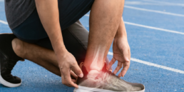
Do you experience sharp heel pain first thing in the morning? You may be suffering from plantar fasciitis. This inflammation of the plantar fascia affects millions of people and represents one of the most common causes of heel pain.
The plantar fascia is a band of fibrous tissue that connects your heel to your toes. When this tissue becomes inflamed or irritated, you develop plantar fasciitis. This painful condition particularly affects runners, overweight individuals, and those who stand for long periods.
Understanding this condition is the first step toward recovery. In this article, you’ll discover the characteristic symptoms, main causes, and effective treatments to regain pain-free mobility.
Definition and Anatomy of Plantar Fasciitis
Plantar fasciitis is an inflammation of the plantar fascia, the fibrous band that connects your heel to your toes. This condition accounts for 15% of foot pain consultations among active adults.
The Plantar Fascia: Structure and Function
The plantar fascia forms an aponeurosis (fibrous membrane that covers or connects muscles) 4-5 millimeters thick that extends under your foot. This fibrous structure originates at the medial tubercle of the calcaneus and divides into 5 bands that attach to the base of each toe.

It performs 3 essential functions:
- Supports the medial longitudinal arch of the foot
- Absorbs shock (up to 2.5 times your body weight)
- Propels the foot during the push-off phase
This aponeurosis works like a biomechanical spring. It stretches 9-12% under normal conditions and stores elastic energy to release during propulsion.
Mechanism of Plantar Fasciitis
Plantar fasciitis develops when repeated microtrauma exceeds your fascia’s regeneration capacity. These microtears appear primarily at the calcaneal insertion, the area subjected to maximum stress.
The inflammatory process follows 3 distinct phases:
| Phase | Duration | Characteristics |
| Acute | 0-6 weeks | Active inflammation, sharp pain |
| Subacute | 6-12 weeks | Decreased inflammation, onset of fibrosis |
| Chronic | >12 weeks | Tissue degeneration, thickening |
Your fascia progressively thickens, increasing from 4mm to 7-10mm in the chronic phase. Collagen fibers lose their parallel organization and adopt a disorganized arrangement. This structural disorganization decreases the tissue’s mechanical resistance and perpetuates the microtear cycle.
Main Causes of Plantar Fasciitis
Plantar fasciitis results from multiple factors that exert excessive tension. These causes are divided into two main categories: biomechanical factors and those related to your lifestyle.
Biomechanical Risk Factors
Your foot anatomy directly influences the development of plantar fasciitis. Flat feet increase your risk of developing this condition by 2.2 times.
High-arched feet also present an increased risk. Your elevated plantar arch concentrates forces on restricted areas of the fascia, creating tension points up to 3 times higher than normal.
Excessive pronation constitutes a major factor. Your foot rolls inward more than 15 degrees during walking. This movement stretches the plantar fascia 10 to 12% beyond its normal length.
Achilles tendon tightness affects 83% of patients with plantar fasciitis. A short Achilles tendon limits dorsiflexion of your ankle to less than 10 degrees. This restriction increases tension on the plantar fascia by 25%.
Lower limb length discrepancies greater than 5 millimeters modify your gait. The longer leg bears an additional load of 20%.
Lifestyle-Related Factors
Your body weight exerts direct pressure on your plantar fascia. A BMI above 30 multiplies your risk of plantar fasciitis by 5.6. Each additional kilogram increases the load on your feet by 4 to 6 kilograms.
Your physical activities influence the onset of fasciitis. Running generates impact forces equivalent to 2.5 times your body weight. Runners covering more than 40 kilometers per week have a 30% increased risk.
Your profession plays a determining role. Occupations requiring prolonged standing (teachers, nurses, workers) expose you to a risk 3.6 times higher. Standing for more than 4 consecutive hours progressively overloads the plantar fascia.
Your inappropriate footwear worsens the situation. Heels higher than 5 centimeters shorten your Achilles tendon by 13%. Worn shoes lose 45% of their shock absorption capacity after 500 kilometers of use.
Age constitutes a non-modifiable factor. Between 40 and 60 years, your plantar fascia loses 20% of its elasticity. The heel fat pad thins by 30%, reducing natural shock absorption.
Symptoms and Diagnosis
Plantar fasciitis manifests through distinctive signs that allow early identification. Recognizing these symptoms and obtaining an accurate diagnosis constitute the first steps toward effective treatment.
Characteristic Signs
Heel pain represents the primary symptom of plantar fasciitis. This pain is precisely localized under the heel, near the calcaneal bone insertion. It is characterized by maximum intensity during the first steps in the morning or after a prolonged period of inactivity.
Painful manifestations follow a typical pattern:
- Burning sensation or stabbing feeling under the heel
- Progressive decrease in pain after 10-15 minutes of walking
- Reappearance of pain after prolonged standing
- Intensification of symptoms after exercise rather than during
Inflammation also causes morning stiffness of the foot. This stiffness particularly affects ankle dorsiflexion (upward movement of the foot toward the leg) and generally disappears after a few minutes of mobilization.
Secondary signs frequently accompany the main pain:
- Mild to moderate heel swelling
- Tenderness to palpation of the medial plantar area
- Gait modification to avoid bearing weight on the painful heel
- Increased tension in the calf and Achilles tendon
Diagnostic Examinations and Tests
The diagnosis of plantar fasciitis is primarily based on a clinical evaluation performed by a healthcare professional. This assessment typically includes a detailed review of your symptoms as well as a thorough physical examination of the foot.
The clinical examination includes several specific tests:
- Passive dorsiflexion test: pain reproduction
- Heel palpation: identification of the precise painful point
- Windlass test: evaluation of tension during toe extension
- Joint mobility analysis: verification of ankle range of motion
Imaging examinations complement the clinical diagnosis in certain cases:
| Type of examination | Usefulness | Frequency of use |
| X-ray | Detects calcaneal spurs (pointed bony growth on the heel) | 30% of cases |
| Ultrasound | Measures fascia thickening (>4mm) | 45% of cases |
| MRI | Evaluates complex tissue lesions | 10% of cases |
Ultrasound constitutes the examination of choice to confirm the diagnosis. It reveals thickening greater than 4 millimeters and allows exclusion of other pathologies such as partial ruptures or cysts.
Complementary analyses allow elimination of other causes of heel pain:
- Tarsal tunnel syndrome
- Calcaneal stress fracture
- Plantar fat pad atrophy
- Peripheral neuropathy
Differential diagnosis relies on the precise location of the pain and symptom characteristics. Plantar fasciitis is distinguished by its characteristic morning pain and specific location at the fascia insertion on the calcaneum.
Prevention and Practical Advice
Prevention of plantar fasciitis is based on concrete actions that reduce the tension exerted. These preventive strategies decrease the risk of developing this painful inflammation by 40 to 60% according to recent clinical studies.
Consult our dedicated article regarding exercises to apply in case of plantar fasciitis: https://laboratoireevo.com/en/relieve-plantar-fasciitis-with-3-exercises/
Exercises and Stretches
Daily stretching constitutes the first line of defense against plantar fasciitis. Your stretching routine targets three anatomical zones: the plantar fascia, the calf, and the Achilles tendon.
Plantar fascia stretch: sitting on a chair, place your affected foot on your opposite knee. Grasp your toes and gently pull them toward your shin for 30 seconds. Repeat this exercise 3 times a day.
Calf stretch against a wall: place your hands against a wall and step your affected leg back. Keep your heel on the ground and lean forward until you feel a stretch in your calf. Hold for 30 seconds and repeat 3 times.
Tennis ball rolling: roll a tennis ball under your arch for 2 minutes. This exercise massages the fascia and improves its flexibility.
Muscle strengthening: Pick up a towel with your toes or walk on your tiptoes for 30 seconds. These exercises strengthen the intrinsic foot muscles that support your plantar arch.
| Exercise | Duration | Frequency | Targeted area |
| Toe work | 30 seconds | 3x/day | Plantar fascia |
| Calf work | 30 seconds | 3x/day | Calf and Achilles tendon |
| Ball rolling | 2 minutes | 2x/day | Plantar arch |
| Plantar flexion | 15 repetitions | 2x/day | Intrinsic muscles |
Choosing Appropriate Footwear
Your shoes play a determining role in preventing plantar fasciitis. Inappropriate footwear increases tension on the fascia by 25 to 35% during walking.
Essential characteristics: look for shoes with a heel of 10 to 25 millimeters, integrated arch support, and a shock-absorbing midsole. The toe box should allow natural movement without compression.
Regular replacement: change your running shoes every 600 to 800 kilometers. Sports shoes should be replaced every 6 to 8 months depending on your frequency of use.
Progressive adaptation: introduce new shoes progressively over 2 weeks. Wear them 1 to 2 hours the first day and gradually increase the duration.
Orthotic insoles: custom orthotics distribute pressure under your foot and correct biomechanical defects. They reduce tension on the plantar fascia by 20 to 30% during walking. Contact the EVO laboratory teams for a personalized assessment of your needs.
House shoes: avoid walking barefoot on hard surfaces. Wear orthopedic sandals or slippers with arch support inside your home.